3D/4D Ultrasound

3D/4D Ultrasound
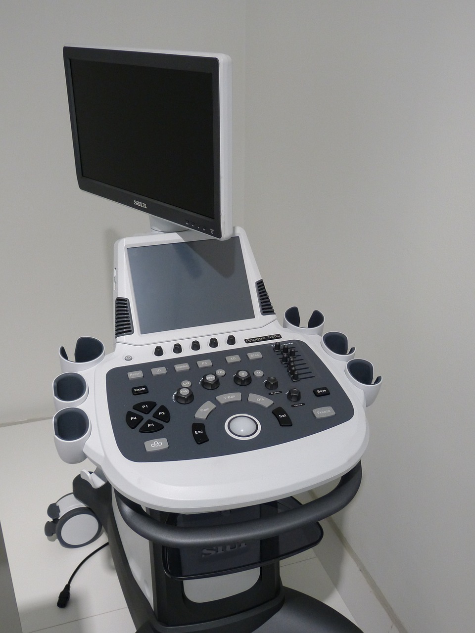
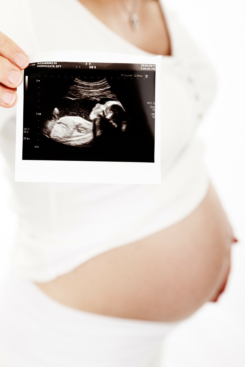
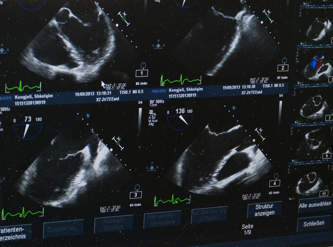
An ultrasound machine creates images that allow various organs in the body to be examined. The machine sends out high-frequency sound waves, which reflect off body structures. A computer receives these reflected waves and uses them to create a picture. Unlike with an x-ray or CT scan, there is no ionizing radiation exposure with this test.
Built on a digital platform, TOP OF THE LINE Voluson BT-13 E-8 utilizes advanced signal processing technology to ensure optimal image quality for high-resolution 2D, volumetric 3D and real-time 4D imaging. Image quality is further enhanced with Harmonic imaging, spectral, color and Doppler imaging,as well as our latest advance – Compound Resolution Imaging. This Machine costs about 4times as much as a routinely used 4D colour Doppler machine and is installed only at a handful of vey reputed diagnostic centres
- Harmonic imaging, spectral, color and Doppler imaging, Compound Resolution Imaging
- High-resolution 2D, volumetric 3D and real-time 4D imaging
- Carotid colour doppler & Transcranial Ultrasound and Doppler in infants
- Peripheral arterial & venous colour Doppler
- Abdominal Colour Doppler
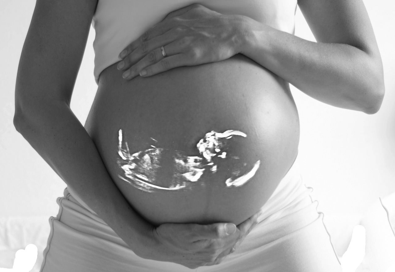
- Pregnancy Colour Doppler
- Musculo-Skeletal Ultrasound.
- Breast Ultrasound
- Neck Ultrasound
Patient Benefits:
- More Information, Better Image Quality
- Faster Examination
- High resolution images for detection of subtle abnormalities.
- Vascular information
What is endovaginal Sonography?
Is dating and weight estimation 100% accurate?
Preparations
- For sonography of Pelvis
- Patient should consume 3 – 4 large glasses of water ½ to 1 hour prior to examination. Urine collects in the bladder over this time. Excessive water should not be consumed as it may lead to vomiting and does not serve the purpose.
- Full bladder permits better evaluation of the urinary bladder itself, the prostate gland and seminal vesicles in males and the uterus, ovaries and adnexa in females.
- Gall Bladder study requires 6 hrs fasting.
- All other examinations do not require any specific preparation.
Pregnancy Sonography
- Full bladder is required upto 10 – 12 weeks of pregnancy. The urinary bladder need not be very full thereafter.
- There are no clothing restrictions. It is advisable to wear something loose and comfortable.
- It is advisable to come 10-15 minutes prior to the appointment to ensure smooth and relaxed procedure.
For KUB Sonography
- No fasting required
- Drink water
- Have full-bladder
For Whole Abdomen Sonography
- 3 – 4 large glasses of water. Urine collects in the bladder takes 1 hour to collect.
- Overnight fasting or 6 hrs fasting required.
For Upper Abdomen Sonography
- 6 hrs fasting required
- Can drink water
Contact us
Call Us
0124-4365459
Email Us
krishna.diagnosticsggn@gmail.com
Our Location
Plot no. 384, Main Market, opposite Raj Bhawan, Block C, Sukhrali, Sector 17, Gurugram, Haryana 122001
